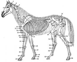
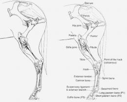
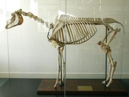
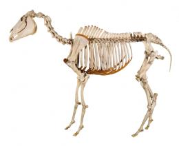
| 1 Incisive bone (premaxillary) | 2 Nasal bone | 3 Maxillary bone | 4 Mandible |
| 5 Orbit | 6 Frontal bone | 7 Temporal fossa | 8 Atlas (first cervical vertebra) |
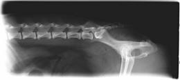
| 9 Axis (2nd cervical vertebra | 10 Cervical vertebrae (7 inc. Atlas and Axis) |
11 Scapular spine | 12 Scapular cartilage |
| 13 Scapula | 14 Thoracic vertebrae (18 of these) |
15 Lumbar vertebrae (6 of these |
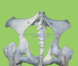
16 Tuber sacrale |
| 17 Sacral vertebrae (5 fused together - sacrum) |
18 Coccygeal vertebrae | 19 Shoulder joint | 20 Ribs (18) |
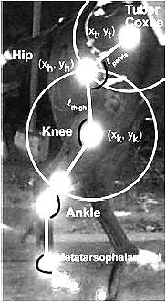
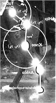
| 21 Costal arch | 22 Tuber coxae | 23 Ilium | 24 Pubis |
| 25 Hip joint | 26 Femur (greater trochanter) |
27 Tuber ischii | 28 Ischium |
| 29 Femur, third trochanter | 30 Femur | 31 Humerla tuberosity, lateral | 32 Humerus |
| 33 Sternum | 34 Olecranon | 35 Costal cartilages | 36 Femoral trochlea |
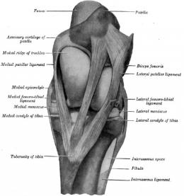
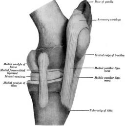
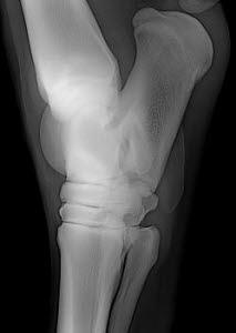
| 37 Stifle joint | 38 Patella | 39 Elbow joint | 40 Ulna |
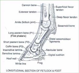
| 41 Radius | 42 CArpus | 43 Metacarpus | 44 Fetlock joint |
| 45 Coffin joint | 46 Accessory carpal bone | 47 Small metarcarpal bone (splint bone) |
48 Proximal sesamoid |
| 49 First phalanx | 50 Distal phalanx | 51 Tibia | 52 Talus (tibial tarsas bone) |
| 53 Small Metatarsal (splint bone) |
54 Metatarsus | 55 Pastern joint | 56 Fibula |
| 57 Calcaneus (fibular tarsal) |
58 Tarsus | 59 Middle Phalanx (2nd phalanx) |
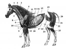
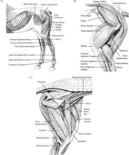
2. Splenius: allows the neck to bend.
3. Multifidus Cervicus (Deep): allows the neck to flex and the head to rotate to the opposite side.
4. Brachiocephalicus: permits the neck to bend, and move the shoulder forward.
5. Trapezius/Rhomboids (Deep): allows the shoulder to raise, and permits the scapula to draw upward, forward and backward.
6. Supraspinatus (Deep): permits the shoulder joint to extend.
7. Infraspinatus (Deep): allows the foreleg to rotate outward.
8. Deltoid: permits the shoulder joint to extend.
9. Tricep: permits the shoulder joint to flex.
10. Bicep and Anterior Pectoral: permits the foreleg to extend.
11. Serratus Thoracis: allows the trunk to be at the proper level when legs are planted.
12. Posterior Pectoral: allows the foreleg to draw backward.
13. Extensor Capri Radialis: permits the foreleg to bend and flex.
14. Latissimus Dorsi: permits lateral bending.
15. Longissimus Dorsi: allows the back to extend, and permits lateral bending.
16. Intercostal: supports the ribcage and aids in respiration.
17. Oblique: allows the hind leg to draw under.
18. Rectus Abdominus: supports the back.
19. Gluteus: allows forward movement and hind end action.
20. Semimembranosus: permits the hock to extend.
21. Semitendinosus: permits the hip and the hock to extend.
22. Bicep Femoris: allows for extension of the hind leg, hip and hock, and bends the stifle.
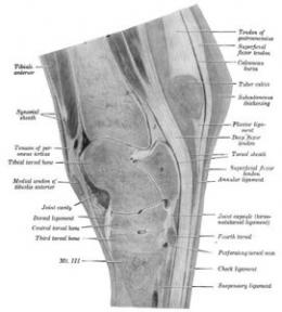
23. Tensor Fascia Latae: allows the stifle to extend and the hip to flex.
24. & Fascia Latae: allows the stifle to extend and the hip to flex.
25. Long Digital Extensor: permits the hind leg to flex.

http://www.red-horse.biz/Hauptpunkte/tendon_injuries.html
http://www.fallight.com/entry/Horse-Conformation
Horse Conformation: Structure, Soundness, and Performance by Equine Research and Sherrie Engler : 사진도 많고 아주 설명이 잘 되어 있다. 입문서, 참고서로 쓰기에 훌륭함.
The Horse in Motion: The Anatomy and Physiology of Equine Locomotion by Sarah Pilliner, Samantha Elmhurst, and Zoe Davies : 이해하기 다소 힘들다. 마필 게이트별 운동원리가 설명되어있음.